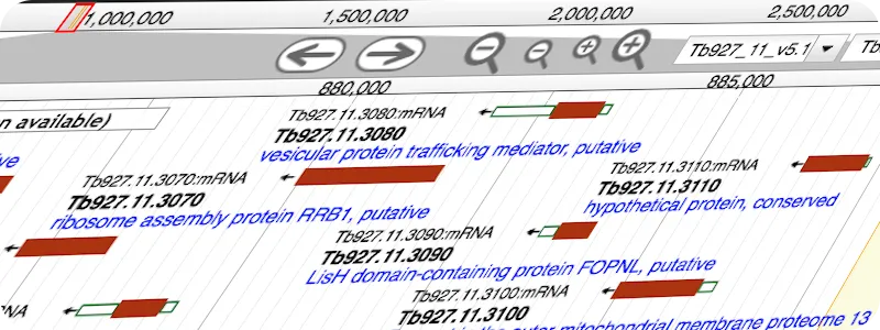
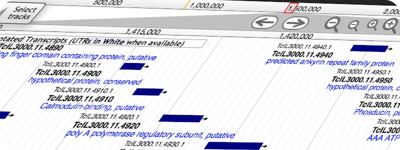
Catarina Gadelha Molecular Cellular Parasitology, Trypanosoma, host-pathogen interactions, infectious diseases, receptors, cell surface membrane

Catarina Gadelha Molecular cellular parasitology, Trypanosom, host-pathogen interactions, infectious diseases, receptors and cell surface membrane biology

gadelha lab

Medicine & Health Sciences

Welcome

Our Lab

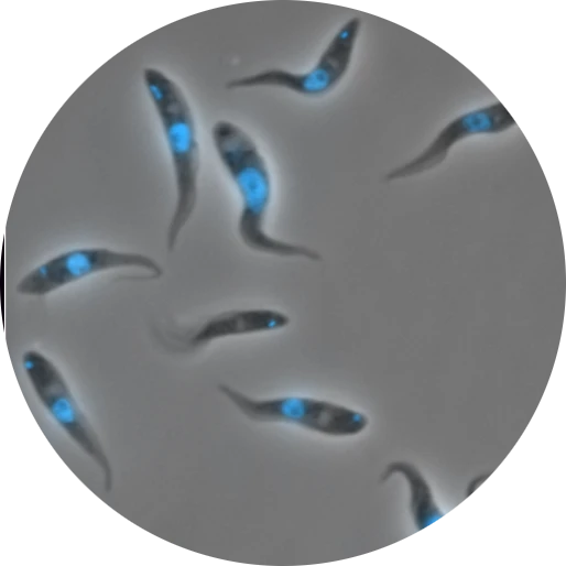
We investigate the structure and function of cell surface membranes in human pathogens, and how these relate to mechanism of disease, evolution and development of treatment. Through studies of cell surface composition and architecture of African trypanosomes, both at the level of molecular targets and of cellular organisation, our work is making significant contributions to membrane biology that is critical for parasitism and to possible exploitation for therapeutic gain.

Catarina Gadelha Molecular cellular parasitology

News

January 2024. Welcome to Rachel, who has recently graduated from Cambridge and joined the group as our new research assistant and lab manager.

April 2023. We have joined Twitter and are excited to share our work and research news with the scientific community. Check us out at @GadelhaLab. The folks in the lab will keep you up to date.

February 2023. This year we are celebrating our 10th lab anniversary! 10 years of experiments, socials, chats, adventures, lots of eppendorfs, tissue culture flasks, tea, coffee and cake. Looking forward to the next decade!

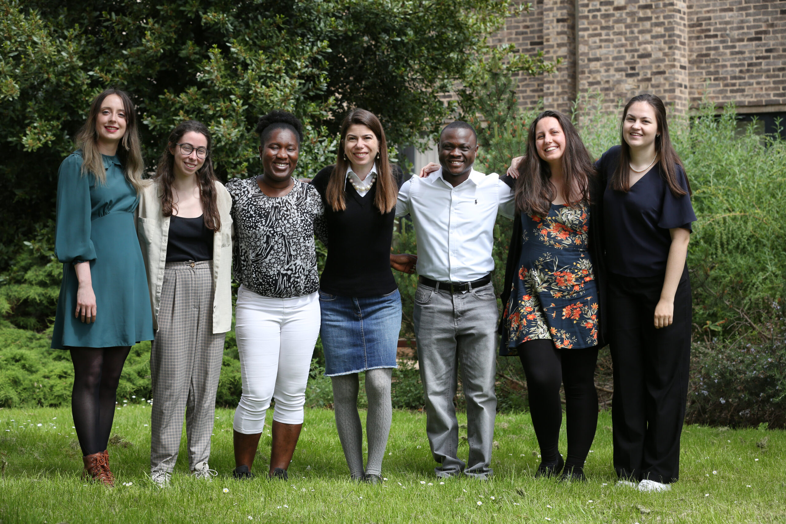
August 2022. Welcome to Majeed, Liz and Ella, who have joined us from Germany, Australia and Ireland.

About Us

Catarina Gadelha Molecular cellular parasitology
Catarina Gadelha

Principal Investigator

Catarina Gadelha, Molecular cellular parasitology ella

Ella Rogerson

Research Assistant

Rachel Angus

Research Assistant

Catarina Gadelha, Molecular cellular parasitology gina2

Georgina Awuah-Mensah

Postdoctoral Fellow

Catarina Gadelha, Molecular cellular parasitology majeed

Majeed Bakari Soale

Postdoctoral Fellow

Catarina Gadelha, Molecular cellular parasitology Liz

Liz King

Postdoctoral Fellow

Research

Cell Surface Biology

For extracellular parasites like African trypanosomes, the cell surface is the primary point of interaction between the organism and the environment – that being the mammalian host or the fly vector. Successful infection and transmission rely on the molecular interactions that take place at this interface. We use cellular, molecular, genomic, and bioinformatic approaches to understand and disrupt the trypanosome cell surface, with an emphasis on mechanism-based research in basic and pre-clinical sciences.

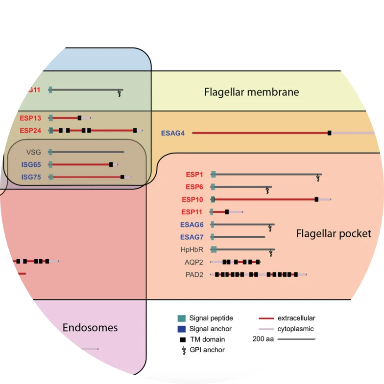
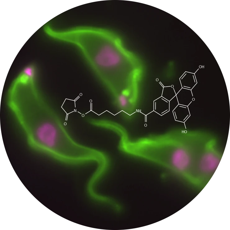
Cell Surface Protein Atlas

Surface membrane proteins are major targets of biomedical research because of their exposure to the extracellular environment and, as such, accessibility for pharmacological intervention. We devised a chemical approach for surface protein capture coupled with quantitative and sensitive mass spectrometry to generate a cell surface protein atlas.

Parasite Genomics

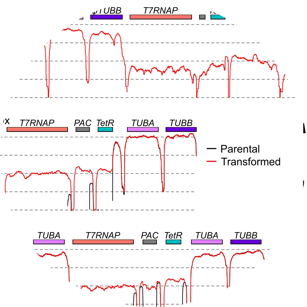
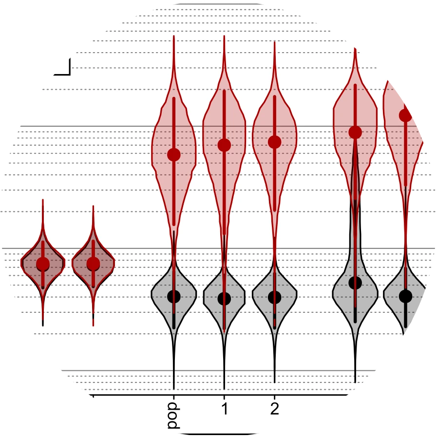
We use genomic methods to interrogate gene fitness in African trypanosome parasites that recapitulate infection dynamics in the host. Our new method (called DRiF-Seq, for Direct RNAi-Fragment Sequencing) robustly measures with high sensitivity the competitive growth rates of >200,000 RNAi mutants (one mutant every ~150 bp across the core diploid genome) in vitro and in vivo.

Emerging Model Systems

Our understanding of eukaryotic cell biology is based on our ability to manipulate experimental systems. Most of our knowledge is centred on a small number of model systems which do not represent the diversity of eukaryotes across the tree of life, nor do they capture all biological phenomena. Advances in genetic technologies and genome sequencing mean that we are now able to address many more fascinating questions in a broader scale, revealing new cell biology and with potential widespread importance.

Publications

Sorting of GPI-anchored proteins at the trypanosome surface is independent of GPI insertion signals.
Miller TH, Schiessler S, Rogerson EM, Gadelha C
The Cell Surface (2024) 12:100131.
PDF
Supplementary Figures
Supplementary Table

A conserved trypanosomatid differentiation regulator controls substrate attachment and morphological development in Trypanosoma congolense.
Silvester E, Szoor B, Ivens A, Awuah-Mensah G, Gadelha C, Wickstead B,  Matthews KR
PLoS Pathogens (2024) 20(2):e1011889.
PDF

A transferrin receptor’s guide to African trypanosomes.
Urbaniak M, Gadelha C.
The Cell Surface (2023) 9:100100
PDF

Reliable, scalable functional genetics in bloodstream-form Trypanosoma congolense in vitro and in vivo.
Awuah-Mensah G, McDonald J, Steketee P et al.
PLoS Pathogens (2021) 17:e1009224.
PDF


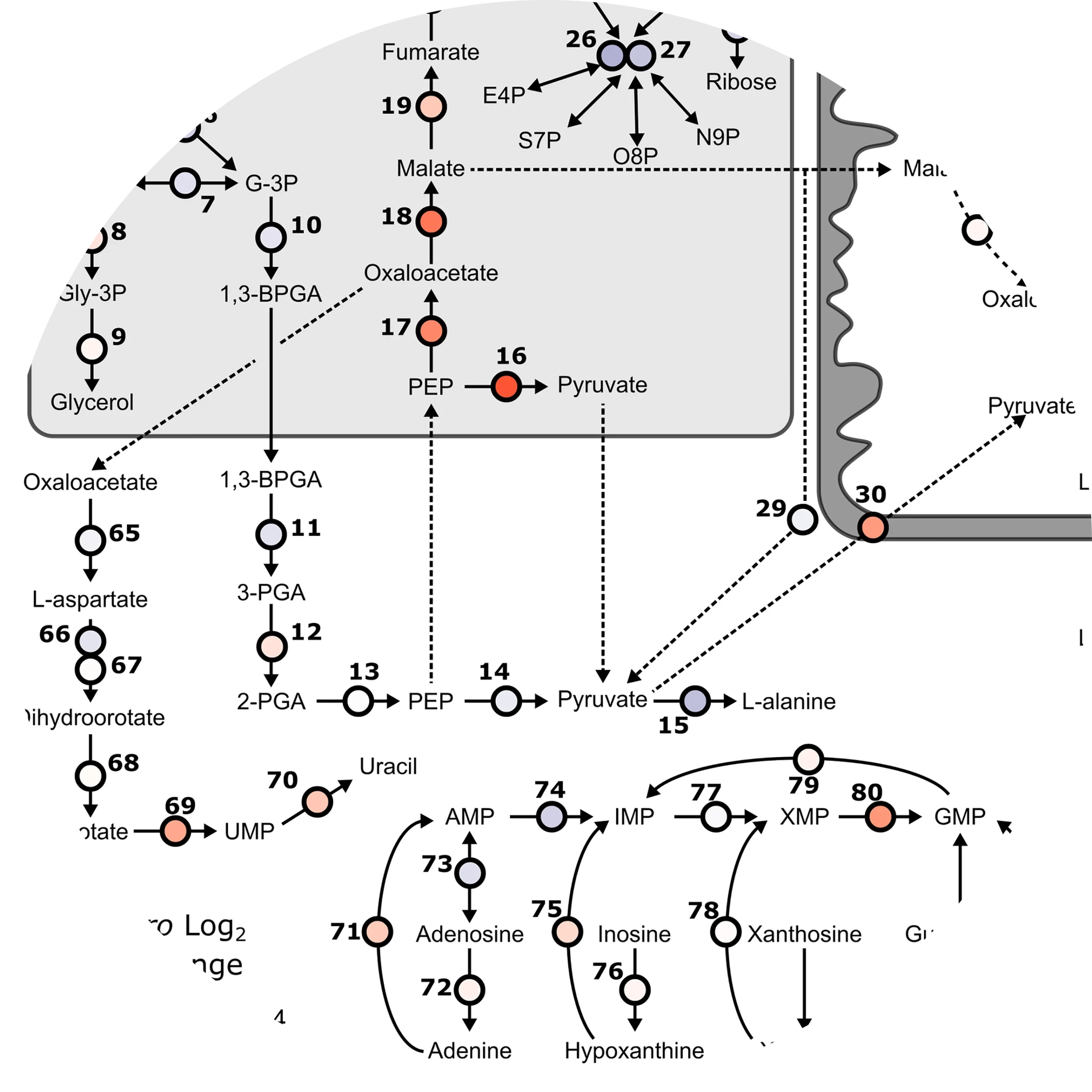
Divergent metabolism between Trypanosoma congolense and Trypanosoma brucei results in differential drug sensitivity.

Steketee P, Dickie E, Iremonger J et al.
PLoS Pathogens (2021) 17:e1009734.
PDF

Suramin exposure alters cellular metabolism and mitochondrial energy production in African trypanosomes.
Zoltner M, Campagnaro G, Taleva G et al.
Journal of Biological Chemistry (2020) 295:8331-8347.
PDF
Supplementary Information

Technical challenges of working with extracellular vesicles.
Ramirez M, Amorim M, Gadelha C, Milic I, Welsh J et al.
Nanoscale (2018) 10:881-906.
PDF

The trypanosome exocyst: a conserved structure revealing a new role in endocytosis.
Boehm C, Obado S, Gadelha C, Kaupisch A, Manna P, Gould G, Munson M, Chait B, Rout M, Field M.
PLoS Pathogens (2017) 13:e1006063.
PDF


Lineage-specific proteins essential for endocytosis in trypanosomes.

Manna P, Obado S, Boehm C, Gadelha C, Sali A, Chait B, Rout M, Field M.
Journal of Cell Science (2017) 130:1379-1392.
PDF
Supplementary Figures

Architecture of a parasite surface: complex targeting mechanisms revealed through proteomics.
Gadelha C, Zhang W, Chamberlain J, Chait B, Wickstead B, Field M.
Molecular and Cellular Proteomics (2015) 14:1911-1926.
PDF
Supplementary Figures
Supplementary Tables


E/ANTH domain proteins participate in AP2-independent clathrin-mediated endocytosis.

Manna P, Gadelha C, Puttick A, Field M.
Journal of Cell Science (2015) 128:2130-2142.
PDF

Proteomic analysis of clathrin interactions in trypanosomes reveals dynamic evolution of endocytosis.
Adung’a V, Gadelha C, Field M.
Traffic (2013) 14:440-457.
PDF
Supplementary Figures


Specificity and function of archaeal DNA replication initiator proteins.

Samson R, Xu Y, Gadelha C, Stone T, Faqiri J, Li D, Qin N, Pu N, Liang Y, She Q, Bell S.
Cell Reports (2013) 3:485-496.
PDF

Identification of ORC1/CDC6-interacting factors in Trypanosoma brucei reveals critical features of origin recognition complex architecture.
Tiengwe C, Marcello L, Farr H, Gadelha C, Burchmore R, Barry JD, Bell SD, McCulloch R.
PLoS ONE (2012) 7:e32674.
PDF
Supplementary Figures

Specializations in a successful parasite: what makes the bloodstream-form African trypanosome so deadly?
Gadelha C, Holden JM, Allison HC, Field MC.
Molecular and Biochemical Parasitology (2011) 179:51-58.
PDF


Discovery of novel intermediate forms redefines the fungal tree of life.

Jones MDM, Forn I, Gadelha C, Egan MJ, Bass D, Massana R, Richards TA.
Nature (2011) 474:200-203.
PDF
Supplementary Figures
Podcast


A novel role for BRCA1 in regulating breast cancer cell spreading and motility.

Coene ED, Gadelha C, White N, Malhas A, Thomas B, Shaw M, Vaux DJ.
The Journal of Cell Biology (2011) 192:497-512.
PDF
Supplementary Figures

In Focus

Basal body movements orchestrate membrane organelle division and cell morphogenesis in Trypanosoma brucei.
Lacomble S, Vaughan S, Gadelha C, Morphew MK, Shaw MK, McIntosh JR, Gull K.
Journal of Cell Science (2010) 123:2884-2891.
PDF
In Focus


Membrane domains and flagellar pocket boundaries are influenced by the cytoskeleton in African trypanosomes.
Gadelha C, Rothery S, Morphew M, McIntosh JR, Severs NJ, Gull K.
Proceedings of the National Academy of Sciences of the U.S.A. (2009) 106:17425-17430.
PDF
Supplementary Figures


Three-dimensional cellular architecture of the flagellar pocket and associated cytoskeleton in trypanosomes revealed by electron microscope tomography.

Lacomble S, Vaughan S, Gadelha C, Morphew MK, Shaw MK, McIntosh JR, Gull K.
Journal of Cell Science (2009) 122:1081-1090.
PDF
In Focus


Flagellar and ciliary beating in trypanosome motility.
Gadelha C, Wickstead B, Gull K.
Cell Motility and the Cytoskeleton (2007) 64:629-643.
PDF


Electron microscopic tomography of the microtubule cytoskeleton and flagellar pocket in African trypanosomes.

Shaw M, Gadelha C, Lacomble S, Morphew M, O’Toole ET, McIntosh JR, Gull K.
Microscopy and Microanalysis (2007) 13:1310-1311.
PDF


Basal body and flagellum mutants reveal a rotational constraint of the central pair microtubules in the axonemes of trypanosomes.

Gadelha C, Wickstead B, McKean PG, Gull K.
Journal of Cell Science (2006) 119:2405-2413.
PDF
In Focus


Evidence for a sliding-resistance at the tip of the trypanosome flagellum.

Woolley D, Gadelha C, Gull K.
Cell Motility and the Cytoskeleton (2006) 63:741-746.
PDF


Cryptic paraflagellar rod in endosymbiont-containing kinetoplastid protozoa.

Gadelha C, Wickstead B, de Souza W, Gull K, Cunha-e-Silva N.
Eukaryotic Cell (2005) 4:516-525.
PDF


Improvement on the visualization of cytoskeletal structures of protozoan parasites using high-resolution field emission scanning electron microscopy (FESEM).

Sant’Anna C, Campanati L, Gadelha C, Lourenço D, Labati-Terra L, Bittencourt-Silvestre J, Benchimol M, Cunha-e-Silva NL, De Souza W.
Histochemistry and Cell Biology (2005) 124:87-95.
PDF


Relationships between the major kinetoplastid paraflagellar rod proteins: a consolidating nomenclature.

Gadelha C, LeBowitz JH, Manning J, Seebeck T, Gull K.
Molecular and Biochemical Parasitology (2004) 136:113-115.
PDF


Further studies on the structural analysis of the cuticle of Litomosoides chagasfilhoi (Nematoda: Filarioidea).

de Moraes Neto AHA, Lanfredi RM, Gadelha C, Cunha-e-Silva NL, Simão RA, Achete C, de Souza W.
Parasitology Research (2003) 89:397-406.
PDF
Resources

We generate research tools to help us address the questions we are most interested in. These tools are freely available to colleagues upon request. Below are some of the plasmids for genetic modification of infective-form Trypanosoma spp. available from us.

In addition to maps, there are annotated GenBank format files containing both sequence and features of each plasmid. These are designed to be used with M. Wayne Davis’s ApE: A Plasmid Editor, which is free, Open Source and runs on Windows/MacOS-X/Linux. GenBank files should also be compatible with your lab’s preferred vector analysis software.

Catarina Gadelha, Molecular cellular parasitology image7A

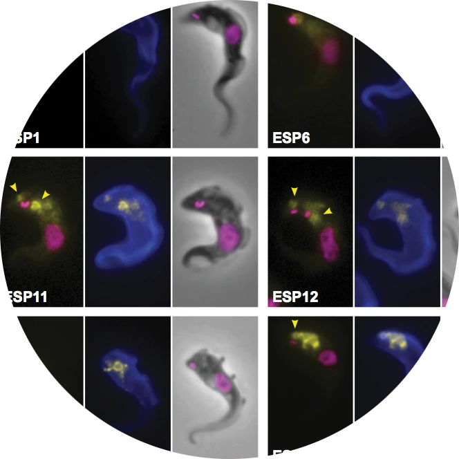
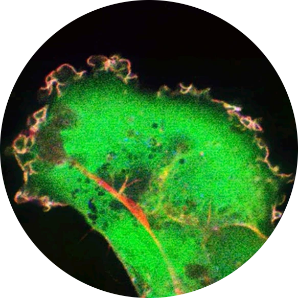
Membrane protein localisation in T.brucei

Catarina Gadelha, Molecular cellular parasitology image7B

Genetic modification of T.congolense

Get in touch

Gadelha Lab

Room D100, Medical School

Queen’s Medical Centre

University of Nottingham

Nottingham, UK NG7 2UH

Lab: +44 115 8230377

Office: +44 115 8230378

We welcome highly motivated individuals with training in chemistry, molecular cell biology, and computational sciences. For information about ongoing research projects, please send your inquiry to catarina.gadelha@nottingham.ac.uk along with your CV.

Gadelha Lab

 © All Rights Reserved